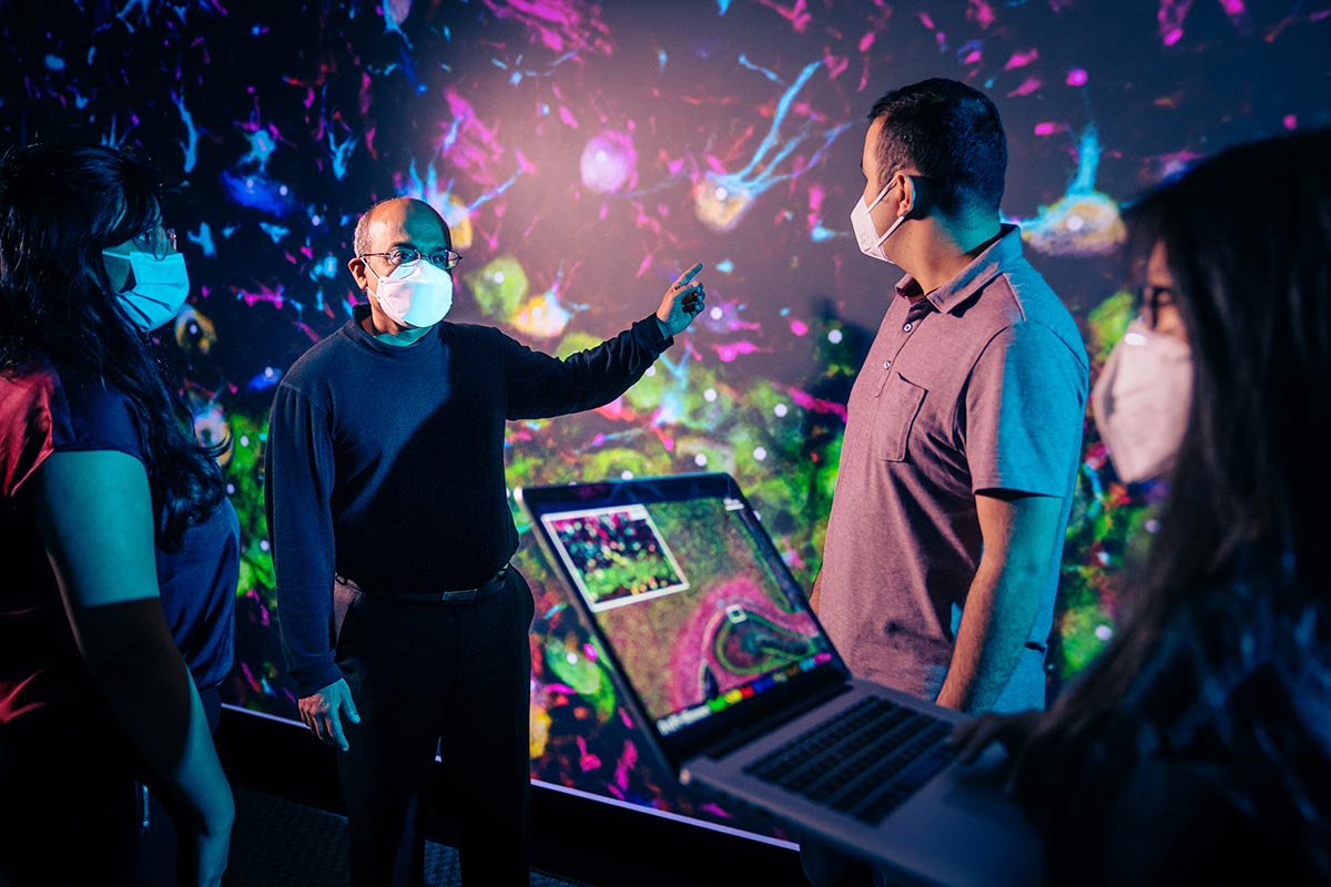
Badri Roysam and students, Aditi Singh, Jahandar Jahanipour and Rebecca Li, review a visualization of brain tissue cells projected in the Research Computing Data Core Visualization Theater at the University of Houston's Hewlett Packard Enterprise Data Science Institute
By Laurie Fickman
Though 40 million concussions are recorded annually, no effective treatment exists for them or for many other brain-related illnesses. In collaboration with Dragan Maric of the National Institutes of Health, Badri Roysam, Hugh Roy and Lillie Cranz Cullen University Professor and Chair of Electrical and Computer Engineering, and his team are working to speed up drug development to treat brain diseases and injuries like concussion by developing new tools.
“We are interested in mapping and profiling unhealthy and drug-treated brain tissue in unprecedented detail to reveal multiple biological processes at once - in context,” said Roysam about his latest paper published in Nature Communications. “This requires the ability to record high-resolution images of brain tissue covering a comprehensive panel of molecular biomarkers, over a large spatial extent, e.g., whole-brain slices, and automated ability to generate quantitative readouts of biomarker expression for all cells.”
At the National Institute of Neurological Disorders and Stroke, Maric developed the innovative imaging technique that can be readily implemented for widespread use with the potential to transform brain studies requiring comprehensive cellular profiling from single and serial slices of brain tissue. Roysam’s lab developed the computational image analysis methods based on deep neural networks. Roysam’s system analyzes the images on the UH supercomputer automatically and can reveal multiple processes at once – the brain injury, effects of the drug being tested and the potential side effects of the drug.
“Compared to existing screening techniques, using iterative immunostaining and computational analysis, our methods are more flexible, scalable and efficient, enabling multiplex imaging and computational analysis of up to 10 – 100 different biomarkers of interest at the same time using direct or indirect IHC immunostaining protocols,” reports Roysam. The new toolkit uses repeated cycles of optimized 10-plex immunostaining with 10-color epifluorescence imaging to accumulate highly enriched image datasets from individual whole-brain slices, from which seamless signal-corrected mosaics are reconstructed and analyzed.
This makes way for more rapid drug development. “We present a direct method that generates readouts for a comprehensive panel of biomarkers from serial whole-brain slices, characterizing all major brain cell types, at scales ranging from subcellular compartments, individual cells, local multi-cellular niches, to whole-brain regions from each slice,” said Roysam.
The open-source toolkit approach is also adaptable to other tissues. Its development can accelerate systems-oriented studies by providing quantitative profiles of all the molecular and cellular players at once, in their detailed spatial context.
“We are efficiently overcoming the fluorescence signal limitations and achieving highly enriched and high-quality source imagery for reliable automated scoring at scale. Our goal is to accelerate system-level studies of normal and pathological brains, and pre-clinical drug studies by enabling targeted and off-target drug effects to be profiled simultaneously, in context, at the cellular scale,” said Roysam.
The team’s work is supported by a $3.19 million grant from the NIH.