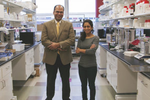
Researchers at the Cullen College’s biomedical engineering Artificial Heart Laboratory (AHL) have recently accomplished a feat valued to researchers across the world: their research was not only chosen for publication in a scholarly journal, but their illustrations were also chosen to serve as the journal’s cover art. Biomedical engineering associate professor Ravi Birla and one of his doctoral students, Nikita Patel, were featured in this month’s American Society for Artificial Internal Organs (ASAIO) journal with an article on regenerating 3D heart muscle.
According to Birla, Patel contributed enormously to both the publication of this article and the associated cover image. Birla said her success in research while still a student illustrates the college’s continued support and development of its doctoral students and their research accomplishments.
The article is called “Engineering 3D Bio-Artificial Heart Muscle: The Acellular Ventricular Extracellular Matrix Model.” It’s a mouthful, but the research is revolutionary for the artificial organ community and represents a significant milestone in the field of tissue fabrication. The AHL is working to create a 3D cardiac patch that will be used to repair, replace and augment lost ventricular function in heart attack patients.
And while she’s still working on obtaining her Ph.D., Patel’s contribution is real, and her explanation of the research illustrates her deep understanding of the issues at hand. “It’s a focus on heart failure,” she explained. “What happens when the heart fails is that some of the tissue dies in one specific section [the left ventricular muscle]. So what they do at the moment to try and fix it is either use heavy pumps, so there will be a lot of pipes and mechanical parts, or they can’t do anything because the tissue is completely dead.” Once heart tissue dies, it is dead forever.
Birla and his research team at the AHL are focused on developing cutting-edge technologies for heart muscle tissue engineering. Their toolbox of tissue and organ models includes 3D heart muscle, blood vessels, tri-leaflet heart valves, tissue engineered heart pumps, bioartificial ventricles and total artificial hearts. The focus of this study was the development of a novel model to bioengineer 3D heart muscle.
In the published study, the patch is designed to either be placed over dead tissue or to replace dead tissue entirely. Once in place, the healthy cardiac cells around the patch begin to regenerate and produce new cardiac tissue to replace the dead, failing tissue.
The patch is a smaller and far less invasive treatment of chronic heart failure than the current method, which includes use of a left ventricular assist device (LVAD). LVADs assist the left ventricular muscle with pumping blood into the aorta, but they are bulky and highly invasive. Pumps and power sources are located outside the body, and tubes connect the pumps to the heart through holes in the abdomen. While patients using an LVAD are able to remain mobile, their way of life is changed greatly after implantation.
“The most invasive part of the LVAD is that once it’s put in, tubes actually have to come out of the chest, but the plan [for the patch] would have it be a one-time open heart surgery, without all the follow up of an LVAD afterward,” Patel said. “It would be all done on the heart without as much interference with the rest of the chest cavity.” Birla added that in the more distant future of organ and tissue bioengineering, the hope is to make procedures like this even less invasive than current open-heart surgeries.
According to Birla, the novelty of this heart muscle model is the utilization of acellular scaffolds to support tissue fabrication. In order to fabricate the acellular scaffolds, whole heart explants were obtained from adult rats. The tissue was essentially scrubbed clean of its current cells to create an acellular total organ. “It’s like having a sponge with nothing on it,” Patel explained.
Then, the excised ventricles were repopulated with neonatal cardiac cells isolated from neonatal rat hearts. “We were wondering if we would be able to get it to behave like a heart muscle would. Can we get it to beat, can it function by itself? And once we put the [neonatal] cells on it and cultured it, that’s exactly what we saw. It’s able to beat by itself as a normal heart muscle would,” Patel said.
In fact, observable muscle contractions occurred on the tissue within 48-72 hours of cell loading, beginning as arrhythmic events but developing to become more synchronized and steady.
Looking ahead, this could mean that a small amount of healthy cells harvested from a donor heart could be placed on a patch and implanted in a failing heart to regenerate new, healthy tissue that would replace the dying tissue.
Birla said he is excited about the future prospects of this technology and its potential utilization in improving the treatment options for heart failure patients. Over the past decade, his research group has developed six completely different and unique platforms to bioengineer 3D heart muscle. These models include the use of self-organization technology, polymeric scaffolds, biodegradable hydrogels, magnetic levitation, in vivo biochambers and acellular scaffolds to support heart muscle formation. Each model provides a unique insight into the complex process of 3D heart muscle formation and function. As this technology is developed further, Birla expects that specific elements from each of the six models will be utilized to support the formation of 3D heart muscle for human use.
You can access the January/February 2015 issue of ASAIO Journal, featuring this research from the AHL as well as the illustration from the lab on the cover, here.
