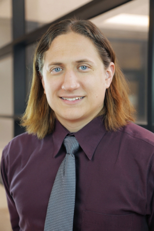
New technologies being developed by a University of Houston researcher to produce three-dimensional models of tissue and whole organ microstructures offer the promise of better diagnosis and treatment for a variety of diseases.
David Mayerich, assistant professor of electrical and computer engineering, received a $984,505 grant from the National Institutes of Health/National Library of Medicine to focus on large-scale reconstruction of microvascular networks.
A wide range of diseases are closely tied to microvascular structure, including several cancers and neurodegenerative diseases.
Mayerich joined the UH Cullen College of Engineering this fall after completing a Beckman Fellowship at the University of Illinois. He specializes in high-performance computing and biomedical imaging and is creating open-source visualization methods, designed to make the information available to biologists for analysis and use in their own research.
His lab focuses on developing new technologies for three-dimensional imaging of whole organs at subcellular resolution. This work is important for biomedical research and ultimately could be valuable at the clinical level, he said.
The work Mayerich proposed under the NIH grant is complementary to work proposed in a $2 million grant that the University received from the Cancer Prevention & Research Institute of Texas to recruit Mayerich to Houston. The CPRIT project involves high-throughput instrumentation and analysis for whole tumor phenotyping.
Both projects were designed to work together, he said. Visualizing large images of tissue microstructure – the subject of the NIH grant – is challenging because biological samples are often densely packed, and high-resolution imaging typically allows only samples of less than half a millimeter to be scanned.
Mayerich has developed much faster methods, allowing data to be collected from whole organs. With the grant, he will use these imaging techniques to convert that data into three-dimensional models, offering researchers and clinicians additional tools.
“The models, we hope would be something researchers and clinicians could use to diagnose and treat disease,” he said.
