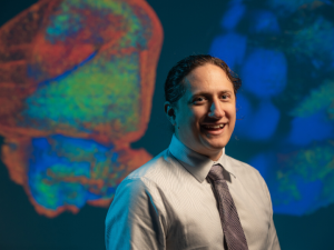
Researchers at the University of Houston last year reported a new imaging method capable of producing fast and inexpensive three-dimensional images, offering the potential to more easily track the progress of conditions such as Alzheimer’s disease and cancer at the cellular level.
David Mayerich, an assistant professor of electrical and computer engineering at UH and corresponding author for a paper describing this work in Scientific Reports, is now at work on Part 2: a software platform to produce searchable digital atlases of whole organs at the cellular level.
He has received a $500,000, five-year CAREER award from the National Science Foundation to pursue the project. The original imaging method grew out of his work with the BRAIN Center, an NSF-supported collaboration between UH and Arizona State University to design, develop and test novel neurotechnologies.
Mayerich describes the project as a Google Maps-style platform, offering both searchability and context for high-resolution 3-D images of whole organs. This platform would allow researchers – and ultimately, clinicians, students and others – to easily search normal and diseased organs to track change over time in animal models and determine how widespread a disease might be at the subcellular level.
“We had a lot of preliminary data from our work at the BRAIN Center, but there wasn’t any software for working with these very large datasets,” he said. “We wanted a platform for doing just that: analyzing massive tissue datasets and make them browsable.”
“We’ve developed this ability to collect massive amounts of data. Now we have to provide the software to make it accessible.”
He said the potential for publicly available whole-organ atlases to affect biomedical research and education is terrific, and routine generation of tissue maps will allow researchers to build detailed models of complex diseases, opening the door to new precision treatments and scalable drug discovery.
Ultimately it could be of use to clinicians and diagnosticians, allowing complete visualization of tissue biopsies instead of more traditional two-dimensional histological slices.
CAREER awards also support educational components, and Mayerich said that in addition to proving useful in educating the next generation of biomedical professionals, he will be working with St. Mary’s, a nearby pre-kindergarten through 6th grade school, to design and test a browsable atlas that can be used for K-12 programs by integrating a virtual reality platform for visualization.
