Struggling to climb out of bed each morning after a restless night’s sleep with stiffness that, at times, slows movement to a standstill is a reality for many Americans. Compounded by uncontrollable muscle movements, basic tasks such as bathing, dressing and eating become arduous efforts that take twice as long as they once did. This is an overly simplistic portrait of the challenges experienced by individuals with Parkinson’s disease.
Approximately 60,000 Americans are diagnosed with the degenerative neurological movement disorder each year, and as many as one million Americans and seven to 10 million people worldwide live with the disease. Common symptoms of Parkinson’s disease, PD, include tremors and other uncontrollable movements, rigidity, slow movement, problems standing, impaired balance and abnormal gait. Patients can also experience cognitive impairment, mood disorders, sleeping problems, slurred speech and difficulty swallowing, among other issues, but the disease is highly individualistic. While one person with the disease might struggle with pushing a button through a shirt’s buttonhole, another might easily sew a missing button on a shirt.
The disorder typically strikes older individuals but approximately 4 percent experience the progressive disease before age 50, according to the Parkinson’s Disease Foundation website. While researchers do not understand the underlying reasons for brain cell death that lead to functional decline in PD patients, and a cure does not exist, they have found ways to treat the disorder.
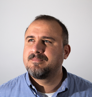
Nuri Firat Ince, assistant professor of biomedical engineering and director of the Clinical Neural Engineering Lab at the UH Cullen College of Engineering, is working to improve one such treatment. With a $350,000, 3-year National Science Foundation grant, he is collaborating with Baylor College of Medicine to develop neural interfaces and signal processing tools for optimization of deep brain stimulation, DBS, a surgical procedure proven to help patients with Parkinson’s disease and other movement disorders.
Ince finds a new window into the human brain for engineers
As a postdoctoral fellow at the University of Minnesota, Ince began composing algorithms for brain-machine interfaces to control communication capabilities for handicapped individuals. To detect and record neuronal activity, he worked mainly with invasive electrodes in nonhuman primates and with noninvasive electroencephalography, EEG, in human subjects.
Limitations of EEG prompted Ince to contact neurosurgeons about potential collaborations for another window on the human brain. His efforts culminated in joint studies of several patients during epilepsy and DBS surgeries in Minnesota.
“This was my first step into the operating room,” Ince said. “Collaborating with neurosurgeons, we placed electrodes on the cortical surface and deep brain during surgeries, and we obtained recordings to understand the pathological neural activity of the brain in PD and epilepsy.”
When Ince joined the Cullen College faculty, his first phone calls were to members of the neurosurgery faculty at the Baylor College of Medicine to explore similar collaborations. For the past two years, Ince and his partners in the Texas Medical Center have studied abnormal neuronal activity in shallow cortical and deep regions of the brain during 35 DBS surgeries, and they have gained ground in optimizing procedural accuracy.
Together, they have collected enough data to publish several joint abstracts and conference papers, which have led to additional funding from Medtronic, a medical device manufacturing company. The interdisciplinary study that coordinates the efforts of Baylor neurologists and neurosurgeons with UH biomedical engineers has provided the first opportunity for a medical resident to work in a UH engineering lab. Similarly, Ince’s research assistant has participated in dozens of DBS surgeries in hospital operating rooms as well as all stages of project development and data analysis in the lab.
“This project provides unique experience for engineering students to see medical problems in the field – in the surgery room – and that provides them with a greater educational environment,” Ince said.
Surgeons open operating rooms to engineers for collaborative research
Ince’s research assistant, Ilknur Telkes, is a doctoral biomedical engineering candidate with a background in neurobiology and neuroinformatics, specifically studying PD in animal models.Before she joined Ince’s lab, Telkes knew little about the engineering techniques that complemented biological sciences. She has since learned about coding, hardware and signal processing as well as detailed operating procedures that coincide with neurosurgeries.
“I took engineering courses, but if you don’t use – in the surgery room – the information, it’s just information, not knowledge,” Telkes said. “This is translational science, from biology to engineering, so we’re trying to understand the neurophysiology of the brain using engineering tools to create devices and techniques for investigation.”
Ince and his team work with Ashwin Viswanathan, a neurosurgeon at Baylor College of Medicine, whose focus is DBS for the treatment of movement disorders and some psychiatric conditions. The team’s objective is to precisely locate the dysfunctional subthalamic nucleus, STN, in the brains of PD patients and to deliver exactly the electrical stimulation necessary to ease problematic symptoms of the disease.
“Historically, we’ve used a trial and error approach in terms of treating patients and finding optimal settings for DBS,” Viswanathan said. “Ince provides a more scientific and quantitative manner of stimulating the brain, so we can record for the abnormal activity, and we can stimulate to treat exactly what’s wrong.”
Novel partnership improves deep brain stimulation outcomes for patients
In 2002, the U.S. Food and Drug Administration approved DBS for PD patients who were unresponsive to medications. During DBS surgery, neurosurgeons implant a battery-powered neurostimulator, generally under the collarbone, that sends electrical impulses up a tiny wire located under the skin to an implanted electrode in the brain. The impulses are delivered chronically to block abnormal electrical signals that cause PD symptoms.
However, available technology and tools cannot target the football-shaped, 4-by-6 millimeter STN with absolute accuracy, so the procedure remains challenging for neurosurgeons. With electrodes, Ince is capturing abnormal firing patterns and oscillations in deep regions of the brain and developing interpretive computer algorithms to help with decision-making.
“Efficacy of DBS depends on accurate placement of the chronic electrode, and a misplaced electrode can cause cognitive side effects,” Ince said. “If clinical decisions are more accurate, then the therapy is better.”
During surgical planning, Viswanathan fuses MRI and CT scan images of the brain with anatomical atlases to determine microelectrode trajectories to his target. Surgeries begin early in the morning, and Ince sets up his computer system in the operating room before the neurosurgeon arrives.
“The most amazing thing is that when we do experiments in the lab, it takes a long time to see outcomes,” Telkes said. “In the OR, we develop algorithms, apply them in the surgery and see results immediately.”
At the beginning of the surgery, Viswanathan positions a neural frame on the skull of the patient to position the electrodes for accurate insertion in the brain. Through a 4-millimeter burr hole in the skull, he pushes three to five microelectrodes 1 millimeter at a time into the brain.
Meanwhile, the electrodes, which are attached to the computer system, record the pathological synchrony in the brain to help determine optimal depth and position for placement of the chronic DBS electrode. The researchers look for excessive and unusual neuronal firing patterns as indicators of their target. Viswanathan then implants the electrode and runs a tiny wire under the skin to the neurostimulator implanted in the chest cavity.
“We are collecting large amounts of data during and after surgery about the patient’s condition, brain function and the effects of stimulation, and we can use that information as a means to develop next-generation technologies,” Viswanathan said. “The partnership between Baylor and UH is one of the strongest we’ve developed, and we look forward to what will happen in the next several years.”
Researchers take deep brain stimulation techniques to the next level
Aside from improved efficacy and fewer side effects for patients, improved DBS precision can extend the life of the battery-powered neurostimulator, which typically lasts three to four years, by targeting specific areas with less electrical stimulation.
“Now with our industry partner, Medtronic, we can take this research to the next stage,” Ince said. “We can move towards a closed loop model where we only stimulate the brain as needed, which is necessary to provide patients with the best possible DBS outcomes.”
Together with Joohi Shahed-Jimenez, a neurologist at Baylor College of Medicine, Ince is extending his DBS research to neurological movement disorders including Tourette’s syndrome, associated with multiple motor tics, and dystonia, characterized by involuntary muscle contractions and abnormal posture.
Ultimately, the goal is to contribute to the broad understanding of the human brain that can lead to effective therapies for these diseases. In addition to determining neuromarkers associated with many cognitive and movement disorders, Ince’s lab uses data collected during surgeries to develop neural decoding algorithms and visual interfaces for developing neuroprosthetics.
“Given outcomes and benefits, I think DBS is going to be an important strategy or treatment technique to suppress the symptoms of Parkinson’s disease and similar movement disorders,” Ince said. “My hope is that in the next few years, we will train engineers who will participate in the surgeries, and who will work closely with neurosurgeons to help with interpretation of signals for electrode placement.”
